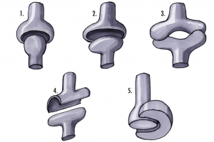
There are a few groups of joint classifications, but in this post, I will focus on those most familiar: the synovial joints. Synovial joints are characterized by the synovial fluid present in the joint capsule that helps to lubricate the joint. The stability of a joint relies on the shape of the joint itself as well as its surrounding structures. This includes the bones that make up the joint, cartilage, ligaments, tendons, and muscles. Cartilage on the bony surfaces that comprise the joint help to increase the surface area of the joint (thereby increasing the stability), and/or cushion the joint. Muscles, tendons, and ligaments help to create support around the joint. The shape of the bones help to dictate which movements it will allow.
Types of Synovial Joints
Within the group of synovial joints, the shape of the articular surfaces of the joint, and the movement allowed at each joint help to further break down joint categories.
1. Ball and Socket Joints: These joints allow for the greatest range of motion. The joint involves a ball fitting into a concave surface. Because these joints allow for more motion, they are at greater risk for instability. Ball and socket joints allow for movement in many planes, and circumduction.
- The shoulder and hip are both ball and socket joints.
2. Condyloid Joints: Allow for flexion, extension, and some lateral movement at the joint. There is also some circumduction that takes place. The circumduction is limited however, because the shape of the joint is oval compared to the more mobile ball and socket joints. Fingers should be able to move in a circular motion, although it is small. Condyloid joints are also referred to as ellipsoidal joints.
- The base of each finger is a condyloid joint.
3. Saddle Joints: These joints are made of two concave and convex surfaces that intersect. Saddle joints allow for flexion, extension, and lateral movement. Saddle joints may present as a condyloid joint, but if you try to move your thumb in a perfect circle, you will notice its movement will not be smooth like the circular movement possible with a finger.
- The base of the thumb is an example of a saddle joint.
4. Hinge Joints: Hinge joints allow for movement in one plane only. Think of a door hinge. It allows the door to open and close, but not move up or down.
- Knees and elbows are some common examples of hinge joints.
5. Pivot Joints: This type of joint allows for rotation. Unlike many other synovial joints, it does not allow for any flexion or extension.
- The first two cervical bones (the atlas & axis) form a pivot joint. The atlas sits on top of the axis and enables a “no” movement of the head (left & right rotation)
Improving stability
Training the muscles around a joint helps to improve its stability. The stronger the muscles, the more control they have over the movements of the joint. A muscle imbalance can also lead to joint laxity.
Post injury, training will generally involve both flexibility and strength training of a joint. The flexibility training is to help regain or improve a joint’s range of motion for daily activities. The strength training is both to help with functional movements alongside building and maintaining joint stability.
For example, I dislocated my shoulder in the Fall of 2014. I performed range of motion and strength exercises to regain my functionality. As I progressed, I was able to do exercises through a wider range of motion to continue improving my stability. The exercises also involved many directions of movement. Training my shoulder this way absolutely brought up the stability (and is still helping to keep it stable). Clearly it will always be at risk for some instability injuries, but training it safely helps me to avoid such instances!
Conclusion
In my last blog post, I explored hypermobility of joints. Overstretching a joint can decrease its overall stability, and overtraining a joint without stretching can decrease its range of motion. There is always a happy medium between stretching and training the muscles for your best function. As always, make sure you stretch what you strengthen and strengthen what you stretch!
Adrienne Lema BPE Student
References:
1. Classification of Joints on the Basis of Structure and Function. Boundless Biology. Boundless, (21 Jul. 2015). Retrieved 30 Jul. 2015 from https://www.boundless.com/biology/textbooks/boundless-biology-textbook/the-musculoskeletal-system-38/joints-and-skeletal-movement-217/classification-of-joints-on-the-basis-of-structure-and-function-820-12063/
2. Joints of the Skeleton, http://www.argosymedical.com/flash/synovial_joints/landing.html , Retrieved from http://legacy.owensboro.kctcs.edu/gcaplan/anat/Notes/API%20Notes%20I%20Types%20of%20Joints.htm
Image References:
1. Joints, Gelenke Zeichnung, (2005) Retrieved from https://en.wikipedia.org/wiki/Ball_and_socket_joint#/media/File:Gelenke_Zeichnung01.jpg
2. Lema, Adrienne (2014)
3. Lema, Adrienne (2014)
4. Lema, Adrienne (2014)


